How is beam width measured in ultrasound?
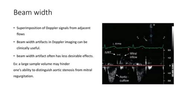
How is beam width measured in ultrasound?
At the transducer, beam width is approximately equal to the width of the transducer. Then, the beam converges to its narrowest width which is half the width of the transducer, at a perpendicular distance from the transducer called the near-zone length (Fig. 2a).
What is beam spread in UT?
Beam spread is a measure of the whole angle from side to side of the main lobe of the sound beam in the far field.
How do you focus an ultrasound beam?
To improve resolution, the ultrasound beam can be focused by using a concave crystal lens or an acoustic lens. Focusing the beam changes the beam’s narrowest point, the focal point, to a narrow area of high resolution, called the focal zone.
What is nearfield length ultrasound?
2.2. Therefore, the near-field length of a beam of ultrasound is related to the diameter (area) of the piezoelectric plate and the speed and frequency of the ultrasound propagation in the medium. At a certain frequency and speed, the larger the diameter, the longer the near-field length.
What is the formula of ultrasound?
Abdominal scans may use a 7-MHz frequency, and the speed of sound in tissue is about 1540 m/s—so the wavelength limit to detail would be λ=vwf=1540 m/s7×106 Hz=0.22 mm λ = v w f = 1540 m/s 7 × 10 6 Hz = 0.22 mm .
What is ultrasound sector width?
Sector width: Selecting the sector width is always a trade-off between the field of view on the one hand and frame rate and image resolution on the other. Therefore, keep sector width at a minimum. Imaging frequency: Most transducers permit the investigator to change the imaging frequency to adapt to individual needs.
How do you calculate beam spread?
A simple calculation to figure out beam spread is to multiply the angle of the beam by the distance.
How do you calculate divergence of a beam?
One may also simply measure the beam intensity profile at a location far away from the beam waist, where the beam radius is much larger than its value at the beam waist. The beam divergence angle may then be approximated by the measured beam radius divided by the distance from the beam waist.
What is beam profile in ultrasound?
The ultrasound beam profile simulation provides the calculated transmitted ultrasound pressure field under certain excitation for a given transducer aperture in a number of different geometrical configurations, including circular elements (flat and concave piston), rectangular elements, linear arrays, convex arrays.
What is an ultrasound beam?
The area through which the sound energy emitted from the ultrasound transducer travels is known as the ultrasound beam. The beam is three-dimensional and is symmetrical around its central axis. Most diagnostic applications, however, use pulsed sound, where the output is a series of short pulses of sound.
How is ultrasound power calculated?
Power of ultrasound is defined as the rate of energy transfer and is measured in Watts. Wavelength is defined as the length of a single cycle. Propagation speed in human soft tissue is on average 1540 m/s. Pulse Duration (msec) = # of cycles x period (msec).