What is a contrast enhanced CT scan?
What is a contrast enhanced CT scan?
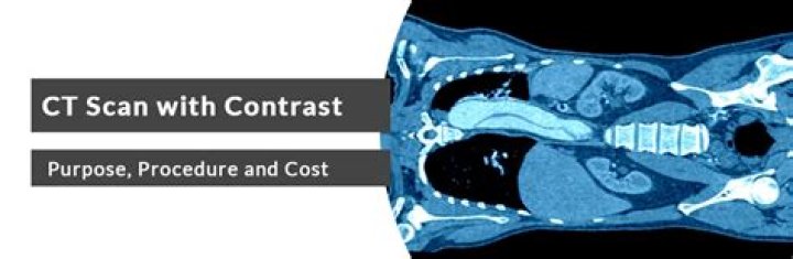
Contrast CT, or contrast enhanced computed tomography (CECT), is X-ray computed tomography (CT) using radiocontrast. Radiocontrasts for X-ray CT are generally iodine-based types. This is useful to highlight structures such as blood vessels that otherwise would be difficult to delineate from their surroundings.
What is the difference between contrast and non contrast CT scan?
CT scans may be done with or without “contrast.” Contrast refers to a substance taken by mouth or injected into an intravenous (IV) line that causes the particular organ or tissue under study to be seen more clearly. Contrast examinations may require you to fast for a certain period of time before the procedure.
What contrasts are used for CT scans?
There are several types of contrast materials: Iodine-based and barium-sulfate compounds are used in x-ray and computed tomography (CT) imaging exams.
Is CT scan contrast harmful?
The IV type: For CT scans, the IV contrast dye we use is iodine-based. It’s safe for most people, but rarely can cause kidney problems in patients who have pre-existing kidney issues, diabetes, or high blood pressure.
When do you use IV contrast CT?
IV contrast is used in brain CT when performing a CT angiogram (or venogram) or for evaluating an abscess or malignancy. In general, workups start with a non-contrast brain CT study and then may progress to MRI or contrast enhanced CT when necessary.
Is CT contrast safe?
What is IV contrast used for?
Benefits of IV Contrast The use of IV contrast greatly improves the accuracy of the examination and assists excluding many life threatening conditions, such as cancer. IV contrast is mainly used to highlight differences between soft tissues which would otherwise look the same.
What is contrast imaging?
Imaging Experience and Advanced Technology Contrast radiography is a method of studying organs using X-rays and the administration of a special dye, called a contrast medium. This test allows the radiologist to evaluate structures that are not clearly evident on conventional X-ray exams.