What is medical imaging used for?
.
In respect to this, what is the purpose of medical imaging?
Medical imaging seeks to reveal internal structures hidden by the skin and bones, as well as to diagnose and treat disease. Medical imaging also establishes a database of normal anatomy and physiology to make it possible to identify abnormalities.
Likewise, is medical imaging the same as radiology? Radiology is a branch of medicine that uses imaging technology to diagnose and treat disease. Radiology may be divided into two different areas, diagnostic radiology and interventional radiology. Doctors who specialize in radiology are called radiologists.
Simply so, what is imaging in medical terms?
Medical imaging refers to techniques and processes used to create images of various parts of the human body for diagnostic and treatment purposes within digital health. The term, medical imaging, includes various radiological imaging techniques such as: X-ray radiography. Fluoroscopy. Magnetic resonance imaging (MRI)
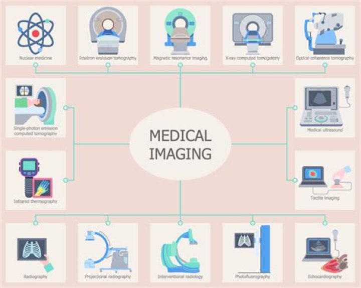
How does each type of medical imaging work?
Other types of medical imaging are magnetic resonance imaging (MRI) and ultrasound imaging. Unlike conventional X-ray, CT and Molecular Imaging, MRI and ultrasound operate without ionizing radiation . Diagnostic ultrasound systems use high-frequency sound waves to produce images of soft tissue and internal body organs.
Related Question AnswersHow long is school for medical imaging?
Certificate or diploma: Certificate programs in medical imaging are typically available through community colleges or vocational schools, and can be completed in anywhere from six months to a year.What are the different types of medical imaging?
There are many different types of imaging, such as X-rays, CT (computed tomography) scans, MRI (magnetic resonance imaging) and ultrasound.What is medical imaging devices?
Tomography is the imaging by sections or sectioning. The main such methods in medical imaging are: Positron emission tomography (PET) also used in conjunction with computed tomography, PET-CT, and magnetic resonance imaging PET-MRI.What is medical imaging salary?
The median salary for this profession was $59,520 in 2018, the BLS reported. The highest paid 10% earned more than $86,350. All diagnostic imaging technologists must be experts in the anatomy they are imaging as well as the technology they are using to create those images.What is the most common form of medical imaging?
The Most Common Types of Medical Imaging- X-Rays. X-rays are among the most commonly used forms of medical imaging.
- MRI. Magnetic resonance imaging, more commonly referred to as an MRI, involves using a magnetic field and radio waves to create highly detailed images of the organs and the tissues inside the body.
- CT Scans.
What happens in an MRI scan?
What happens during an MRI scan? During an MRI scan, you lie on a flat bed that's moved into the scanner. Depending on the part of your body being scanned, you'll be moved into the scanner either head first or feet first. At certain times during the scan, the scanner will make loud tapping noises.What is considered advanced imaging?
MIPPA defines advanced diagnostic imaging procedures as including diagnostic MRI, CT, and nuclear medicine imaging such as positron emission tomography (PET). The accreditation requirements only apply to the technical component of diagnostic imaging and not a physician's interpretation.How do imaging devices work?
A special camera or imaging device detects radioactive emissions from the radiotracer. The camera or device produces pictures and provides molecular information. Many centers superimpose nuclear medicine images with computed tomography (CT) or magnetic resonance imaging (MRI) to produce special views.Is radiology a doctor?
Radiologists are medical doctors that specialize in diagnosing and treating injuries and diseases using medical imaging (radiology) procedures (exams/tests) such as X-rays, computed tomography (CT), magnetic resonance imaging (MRI), nuclear medicine, positron emission tomography (PET) and ultrasound.Is radiography hard to study?
While radiography is not extremely difficult to learn, there is an immense amount to learn. The volume of information that must be learned and retained is considerable. If you meet the prerequisites for admission to the program, there is no academic reason you should not succeed.What are the types of radiology?
The most common types of diagnostic radiology exams include:- Computed tomography (CT), also known as a computerized axial tomography (CAT) scan, including CT angiography.
- Fluoroscopy, including upper GI and barium enema.
- Magnetic resonance imaging (MRI) and magnetic resonance angiography (MRA)
- Mammography.