What is Schwalbes line
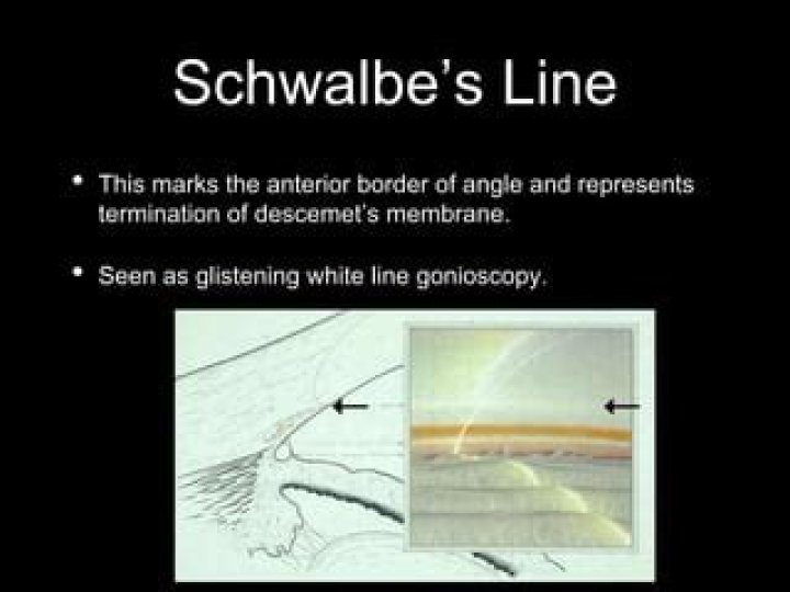
Schwalbe’s line (SL) is often defined as the posterior limbal
Is Schwalbe's line pigmented?
During gonioscopy (where the structures of the eye’s anterior segment are examined), if an abundance of brown pigment is seen at or anterior to Schwalbe’s line, a Sampaolesi line is said to be present.
What is scleral spur?
The scleral spur is a fibrous ring that, on meridional section, appears as a wedge projecting from the inner aspect of the anterior sclera (Figs 3-1 and 3-2). The spur is attached anteriorly to the trabecular meshwork and posteriorly to the sclera and the longitudinal portion of the ciliary muscle.
What is normal angle of eye?
Based on a previous study of anterior segment OCT, the mean anterior chamber angle of healthy normal eyes was 35.9 ± 5.7°.What is scleral sulcus?
In front of eyeball, the sclera is directly continuous with the cornea, the line of union being termed the sclero-corneal junction. This line forms a shallow groove produced by the stronger curvature of the cornea, called the sulcus sclerae.
What is angle recession glaucoma?
Angle recession glaucoma (ARG) is a secondary open angle glaucoma that is associated with ocular trauma. Recession of the anterior chamber angle is a common slit lamp and gonioscopic finding following concussive ocular trauma.
What causes plateau iris?
Plateau iris is caused by a narrowing of the anterior chamber angle due to insertion of the iris anteriorly on the ciliary body or displacement of the ciliary body anteriorly, which in turn alters the position of the peripheral iris in relation to the trabecular meshwork (i.e. placing them in apposition).
How do you find the angle between iris and cornea?
a) AOD 500 (Angel Opening Distance) involves measuring a distance between a point of the cornea which is 500 μm away from the scleral spur and the opposite point of the iris. b) TIA (Trabecular-Iris Angle) involves a direct measurement of the angle.What is open angle glaucoma?
Open-angle glaucoma is the most common form of the disease. The drainage angle formed by the cornea and iris remains open, but the trabecular meshwork is partially blocked. This causes pressure in the eye to gradually increase. This pressure damages the optic nerve.
What is pars plicata?The pars plicata is the portion of the ciliary body that is responsible for producing aqueous humor, the fluid of the anterior chamber. The production of too much aqueous humor, or reabsorption that occurs too slowly, can lead to increases in the pressure within the eye.
Article first time published onWhat is ocular Hypotony?
Hypotony is usually defined as an intraocular pressure (IOP) of 5 mm Hg or less. Low IOP can adversely impact the eye in many ways, including corneal decompensation, accelerated cataract formation, maculopathy, and discomfort. Clinically significant changes occur more frequently as the IOP approaches 0 mm Hg.
How is a scleral buckle done?
Dilation widens your pupil, allowing your doctor to see the back of your eye. Once the conjunctiva (or clear, skin of the eye) is peeled back to expose the white part of the eye (sclera) the buckle is then secured to the white of the eye, behind the eyelids and under the eye muscles. Once in place, it will not move.
What is iris eye?
The colored tissue at the front of the eye that contains the pupil in the center. The iris helps control the size of the pupil to let more or less light into the eye.
What is posterior to the iris?
Posterior chamber: The posterior chamber is between the iris and lens. The lens is behind the iris and is normally clear. Light passes through the pupil to the lens. … Small muscles attached to the lens can change its shape, allowing the eye to focus on objects at varying distances.
What produces conjunctiva?
The conjunctiva helps lubricate the eye by producing mucus and tears, although a smaller volume of tears than the lacrimal gland. It also contributes to immune surveillance and helps to prevent the entrance of microbes into the eye.
What causes Peters anomaly?
The cause of Peters anomaly is unknown; it may be caused by genetic factors (including alterations of several genes , like the FOXC1, PAX6, PITX2, or CYP1B1 genes, environmental factors , or both. The critical event must occur in the first trimester of pregnancy during the formation of the anterior chamber.
Why is gonioscopy important?
Gonioscopy, which enables visualization and magnification of the iridocorneal angle through the use of a gonioscopic lens, “is very important for the diagnosis of glaucoma in terms of our major divisions of glaucoma, open angle and closed angle.
How often should gonioscopy be done?
The AAO’s Preferred Practice Patterns suggests that gonioscopy be repeated periodically and mentions every 1 to 5 years. Repeat testing is indicated when medically necessary for new symptoms, progressive disease, new findings, unreliable prior results, or a change in the treatment plan.
What is the difference between Iridotomy and Iridoplasty?
Currently, opinion is that laser peripheral iridotomy (LPI) alone is not sufficient to prevent disease progression. Laser peripheral iridoplasty (LPIP) is an alternative and effective way of widening the angle recess in eyes that are affected by primary angle closure (PAC).
How is Iridoplasty performed?
In an iridoplasty procedure, a laser beam is applied to the iris (the coloured part of your eye) next to the drainage channel. This causes contraction of the iris, which helps to open up the drainage channels. As a result, the aqueous humour can flow through the channels better.
What is indentation Gonioscopy?
Indentation gonioscopy is a strategy that helps determine whether angle closure is the result of the iris being in apposition (i.e., just touching the angle) or the result of the iris actually being stuck on the angle, via synechiae. Indentation gonioscopy is also a great tool for diagnosing plateau iris.
How is angle recession treated?
Your doctor may prescribe medications, suggest laser treatment, or consider surgery. Medication: Prescription eye drops are usually the first step in treatment. For angle recession glaucoma, your doctor may prescribe a drop that causes your eye to make less fluid. That helps lower pressure.
What does your vision look like with glaucoma?
According to a study published in The American Journal of the Medical Sciences, the most common visual symptoms reported by patients with glaucoma are as follows: Needing more light. Blurry vision. Seeing glare.
What kind of trauma causes glaucoma?
As a result of an immediate injury, traumatic glaucoma is most commonly caused by blunt trauma, which is an injury that doesn’t penetrate the eye, such as a blow to the head or an injury directly on the eye. The most common cause is from sports-related injuries, such as baseball or boxing.
What medications should be avoided with open angle glaucoma?
Steroids are the most important open angle glaucoma medication to avoid. Steroid usages can cause permanent blockage of the eye’s drainage system.
Is eye pressure of 50 high?
In general, pressures of 20-30 mm Hg usually cause damage over several years, but pressures of 40-50 mm Hg can cause rapid visual loss and also precipitate retinovascular occlusion.
What foods to avoid if you have glaucoma?
- Caffeine. Some studies suggest caffeine increases intraocular pressure, cholesterol levels, and blood pressure. …
- Saturated Fats. …
- Trans Fats. …
- Weight-Lifting. …
- Scuba Diving. …
- Bungee Jumping. …
- Yoga.
What is optic nerve head?
The optic disc or optic nerve head is the point of exit for ganglion cell axons leaving the eye. Because there are no rods or cones overlying the optic disc, it corresponds to a small blind spot in each eye.
What is the acute angle?
Acute angles measure less than 90 degrees. Right angles measure 90 degrees. Obtuse angles measure more than 90 degrees.
What is Uveoscleral outflow?
The term uveoscleral outflow refers to the drainage of ocular aqueous humor from the anterior chamber into the anterior chamber angle other than through the trabecular meshwork (Figure). Unlike the trabecular outflow route, the uveoscleral outflow route is not a distinctive pathway with tubes and channels.
What do Zonular fibers do?
The zonular fibers anchor the the equator of the lens and adjacent anterior and posterior surface of the lens to the ciliary body and and ciliary part of the retina. The ciliary epithelial cells of the eye probably synthesize portions of the zonules.