What is the difference between hypoechoic and hyperechoic?
What is the difference between hypoechoic and hyperechoic?
Hypoechoic: Gives off fewer echoes; they are darker than surrounding structures. Examples include lymph nodes and tumors. Hyperechoic: Increased density of sound waves compared to surrounding structures. Examples include bone and fat calcifications.
What does hyperechoic or Isoechoic mean?
Hyperechoic. This term means “lots of echoes.” These areas bounce back many sound waves. They appear as light gray on the ultrasound. Hyperechoic masses are not as dense as hypoechoic ones are. They may contain air, fat, or fluid.
What does hypoechoic mean on ultrasound?
A hypoechoic mass is tissue in the body that’s more dense or solid than usual. This term is used to describe what is seen on an ultrasound scan. Ultrasound uses sound waves that are absorbed by or bounce off of tissues, organs, and muscles. The waves form the black and white image you see on an ultrasound screen.
What is the difference between echogenic and hyperechoic?
In other words, echogenicity is higher when the surface bouncing the sound echo reflects increased sound waves. Tissues that have higher echogenicity are called “hyperechogenic” and are usually represented with lighter colors on images in medical ultrasonography.
What is the normal size of hypoechoic lesion?
The lesions measured from 6 to 20mm (mean 13.1 mm).
What color is hypoechoic?
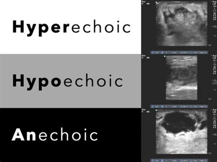
Based on echogenicity, a structure can be characterized as hyperechoic (white on the screen), hypoechoic (gray on the screen) and anechoic (black on the screen) [Figure 1].
What is hyperechoic structure?
Hyperechoic – A relative term that refers to the echoes returning from a structure. Hyperechoic tissues generate a greater echo usually displaying as lighter colors during ultrasound imaging. Hypoechoic – Refers to structures that create weaker echoes such as a fluid.
What is hyperechoic area in uterus?
The term “hyperechoic” is used to describe how the tissue looks during an ultrasound exam. This is a rather nonspecific term meaning that during the test the tissue reflected back an unusually large number of ultrasound echoes.
What percentage of hypoechoic masses are malignant?
In addition increase in vascularity in the hypoechoic mass predicts malignancy about 82% of the time. The ultrasound image below shows an irregular vascularized retroareolar mass, with calcifications. This is very likely to be infiltrating ductal carcinoma and your doctor will recommend a biopsy straight away.
Can fibroids be hyperechoic?
Fibroids may vary in their degree of echogenicity; they can be heterogeneous or hyperechoic, depending on the amount of fibrous tissue and/or calcification. Fibroids may have anechoic components resulting from necrosis.