What kind of MRI is used for hydrocephalus?
What kind of MRI is used for hydrocephalus?
The PC-MRI and/or 3D-SPACE methods are relatively simple for evaluating true CSF flow and determining the obstruction level. Also, these techniques provide additional physiological information. The 3D-SPACE technique seems to be the most efficient and rapid for evaluating hydrocephalus, ETV and the shunt catheter.
What does hydrocephalus look like on MRI?
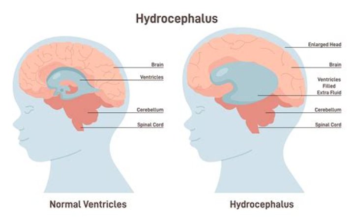
CT/MRI criteria for acute hydrocephalus include the following: Size of both temporal horns is greater than 2 mm, clearly visible. In the absence of hydrocephalus, the temporal horns should be barely visible.
Does hydrocephalus show on MRI?
MRI scans can show enlarged ventricles caused by excess cerebrospinal fluid. They can also be used to identify causes of hydrocephalus or other conditions contributing to the symptoms.
How do you measure NPH?
How is normal pressure hydrocephalus (NPH) diagnosed?
- Imaging tests. A CT scan or MRI of the head is done to look for enlarged ventricles in the brain.
- Cerebrospinal fluid tests. These tests include a spinal tap and external lumbar drainage.
- Gait analysis (walking). This is a timed walk test.
- Neuropsychological testing.
Can lumbar puncture treat hydrocephalus?
A large volume lumbar puncture is a special kind of lumbar puncture (spinal tap) specifically intended to remove 30 to 40 ml of cerebrospinal fluid (CSF) to both assess and temporarily relieve symptoms of hydrocephalus.
How can you distinguish between obstructive and communicating hydrocephalus?
The word “communicating” refers to the fact that CSF can still flow between the ventricles, which remain open. Non-communicating hydrocephalus – also called obstructive hydrocephalus – occurs when the flow of CSF is blocked along one or more of the narrow passages connecting the ventricles.
Can hydrocephalus be misdiagnosed?
The Hydrocephalus Association estimates that nearly 700,000 adults have normal pressure hydrocephalus, but it is often misdiagnosed as Alzheimer’s or Parkinson’s disease. In fact, less than 20 percent of people with the disease are properly diagnosed.
Can a lumbar puncture diagnose hydrocephalus?
Why It Is Performed When a large volume lumbar puncture is used to assess normal pressure hydrocephalus, your doctor will assess your gait and balance both before and after the procedure to see if removal of CSF results in a significant improvement. These tests will help guide treatment decisions.
How do you detect hydrocephalus?
To diagnose hydrocephalus, a neurosurgeon or neurologist takes a thorough medical history and performs a neurological evaluation and physical exam. Doctors then perform an ultrasound, MRI scan, or CT scan to confirm enlargement of the ventricles and determine the cause of the hydrocephalus.