How do you find the landmarks for precordial lead placement?
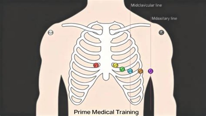
- To locate the space for V1; locate the sternal notch (Angle of Louis) at the second rib and feel down the sternal border until the fourth intercostal space is found.
- Next, V4 should be placed before V3.
- V3 is placed directly between V2 and V4.
- V5 is placed directly between V4 and V6.
.
Hereof, where are the precordial leads placed?
The precordial (chest leads) leads each consist of a positive electrode strategically placed on the chest of the patient.
Likewise, where do electrodes go on ECG? Place the fifth intercostal space at the mid-axillary line. As if drawing a line down from the armpit (mid-axillary line), place the V6 electrode at the fifth intercostal space. Electrodes V4, VA, and V6 should line up horizontally along the fifth intercostal space.
Likewise, where are the 12 leads placed on a patient for an ECG?
To properly record a 12-lead ECG, it is important to have the patient lying comfortably with the wrist close to but not touching the trunk. The limb electrodes should be placed on the right and left wrists and the right and left ankle.
What are the right precordial leads?
The precordial, or chest leads, (V1,V2,V3,V4,V5 and V6) 'observe' the depolarization wave in the frontal plane. Example: V1 is close to the right ventricle and the right atrium. Signals in these areas of the heart have the largest signal in this lead. V6 is the closest to the lateral wall of the left ventricle.
Related Question AnswersWhy is a 12 lead called a 12 lead?
The 12-lead ECG displays, as the name implies, 12 leads which are derived by means of 10 electrodes. Three of these leads are easy to understand, since they are simply the result of comparing electrical potentials recorded by two electrodes; one electrode is exploring, while the other is a reference electrode.What does aVF stand for?
aVR means augmented Vector Right; the positive electrode is on the right shoulder. aVL means augmented Vector Left; the positive electrode is on the left shoulder. aVF means augmented Vector Foot; the positive electrode is on the foot.How do precordial leads work?
As a result of the "location" of the ground lead being in the center of the chest, the precordial leads measure electrical activity that is moving in a front-back direction and/or a right-left direction. Unlike the limb leads, they do not measure any signals in the up-down (head-toe) direction.Why does a 12 lead have 10 leads?
The 12 Lead Groups. A lead is a glimpse of the electrical activity of the heart from a particular angle. In 12-lead ECG, there are 10 electrodes providing 12 perspectives of the heart's activity using different angles through two electrical planes - vertical and horizontal planes.What does axis deviation tell us about the heart?
In electrocardiography, left axis deviation (LAD) is a condition wherein the mean electrical axis of ventricular contraction of the heart lies in a frontal plane direction between −30° and −90°. This is reflected by a QRS complex positive in lead I and negative in leads aVF and II.What is aVR lead?
CLINICAL UTILITY OF LEAD aVR The lead aVR is oriented to 'look' at the right upper side of the heart, and can provide specific information about the right ventricle outflow tract and basal part of the septum (10).What does the V stand for on ECG leads?
The V stands for vector here. In aVR, the left leg lead and left shoulder lead BOTH provide the negative pole for the EKG while the right shoulder is positive. The augmented vector leads require THREE poles to work: two negative and one positive.What is a normal ECG reading?
Normal range 120 – 200 ms (3 – 5 small squares on ECG paper). Normal range up to 120 ms (3 small squares on ECG paper). QT interval (measured from first deflection of QRS complex to end of T wave at isoelectric line). Normal range up to 440 ms (though varies with heart rate and may be slightly longer in females)Where does v1 lead go?
V1 is placed to the right of the sternal border, and V2 is placed at the left of the sternal border. Next, V4 should be placed before V3. V4 should be placed in the fifth intercostal space in the midclavicular line (as if drawing a line downwards from the centre of the patient's clavicle).What is happening in the heart in a normal ECG trace?
An ECG is performed by placing electrodes on the skin overlying the heart. As the electrical impulse moves from the atria, which are the top two chambers, to the ventricles down below, the voltage measurement between the electrodes varies, and this produces a graph of how your heart is performing.How do you read an ECG?
How to Read an ECG- Step 1 – Heart rate.
- Step 2 – Heart rhythm.
- Step 3 – Cardiac axis.
- Step 4 – P-waves.
- Step 5 – P-R interval.
- Step 6 – QRS complex.
- Step 7 – ST segment.
- Step 8 – T waves.
How is ECG procedure done?
An EKG is quick, painless, and harmless. After you change into a gown, a technician attaches 12 to 15 soft electrodes with a gel to your chest, arms, and legs. The technician may have to shave small areas to ensure the electrodes stick properly to your skin. Each electrode is about the size of a quarter.Which precordial leads are most commonly used in bedside monitoring?
Lead II and MCL1 (Fig. 5.2) are in common use for bedside monitoring because they most consistently show the p wave.How do you do a 12 lead?
Precordial Lead Placement- To locate the space for V1; locate the sternal notch (Angle of Louis) at the second rib and feel down the sternal border until the fourth intercostal space is found.
- Next, V4 should be placed before V3.
- V3 is placed directly between V2 and V4.
- V5 is placed directly between V4 and V6.
What are bipolar leads?
Bipolar Leads. Well, the 2 leads situated on the right and left wrist (or shoulders), AVr and AVL respectively, and the lead situated on the left ankle (or left lower abdomen) AVf, make up a triangle, known as "Einthoven's Triangle". Information gathered between these leads is known as "bipolar".Where do you place a 5 lead ECG?
For a 5-lead system, you'll also place the following:- GREEN.
- RL (right leg), on the lower chest, just above and to the right of the umbilicus.
- BROWN.
- (representing any of the six precordial leads), generally in the V 1 position at the fourth intercostal space, right sternal border.
How do you place a 3 lead ECG electrode?
3 lead Placement (I, II, or III):- RA: red electrode: placed under right clavicle near right shoulder, within the rib cage frame.
- LA: yellow electrode: placed under left clavicle, near left shoulder, within the rib cage frame.
- LL: green electrode: placed on the left side, below pectoral muscles, lower edge of left rib cage.